http://www.fantinlab.unimi.it/
Home
Research
Publications
Collaborations
Research pictures
Members
Lab members
Lab pictures
News
News
Useful links
Contacts
Contacts
Open positions
create your own web page for free
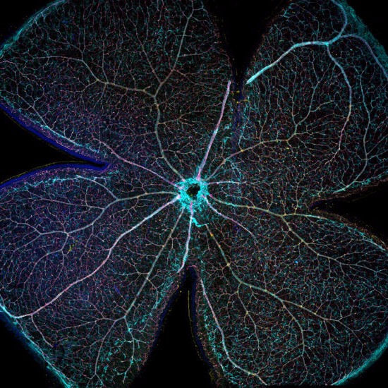
Blood vessels in the retina (author: Veronica Bonalume)
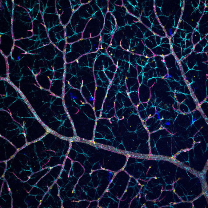
Blood vessels in the retina, close up (author: Veronica Bonalume)
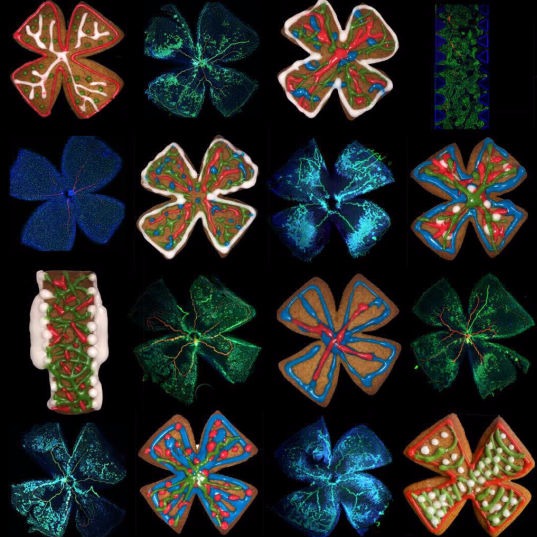
Blood vessels in the retina, spot the biscuit! (author: Veronica Bonalume and Fantin lab)
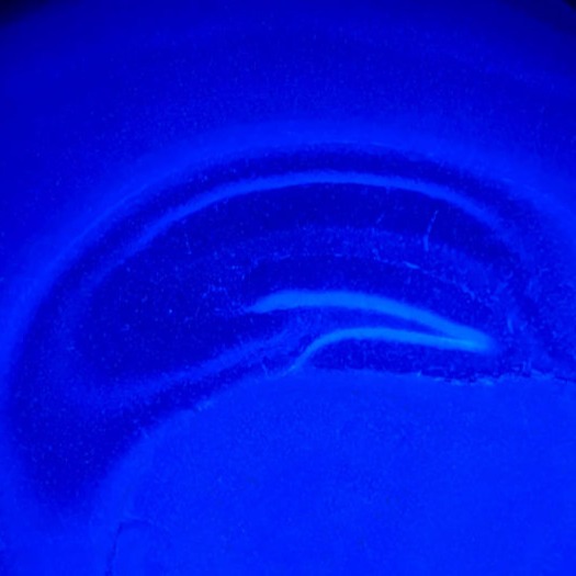
Adult brain hyppocampus (author: Veronica Bonalume)
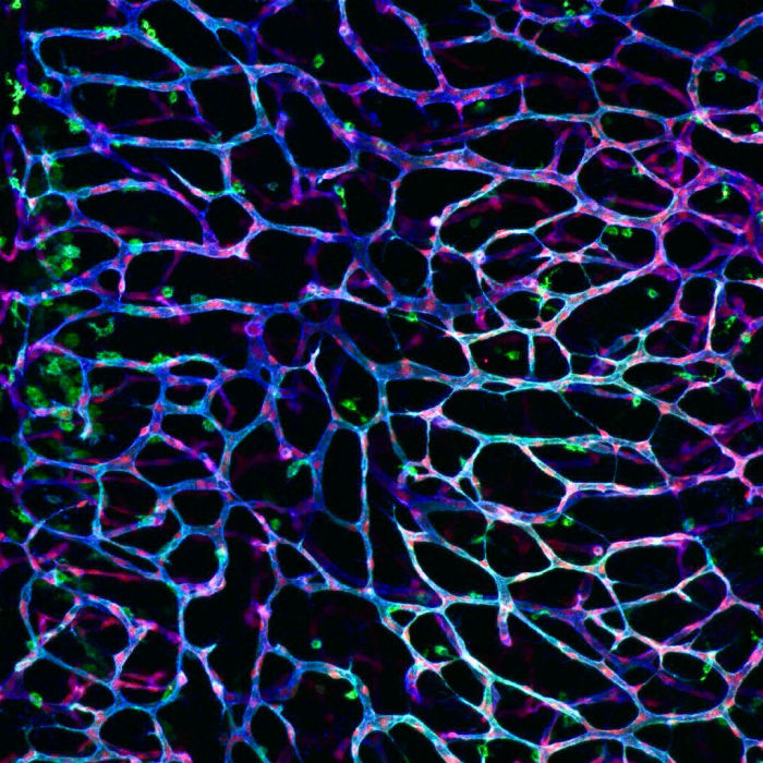
Embryonic brain blood vessels (author: Veronica Bonalume)
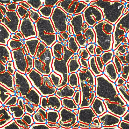
Virtual skelethonization of the blood vessels from the mouse embryo brain acquired with confocal microscopy (author: Alison Domingues)
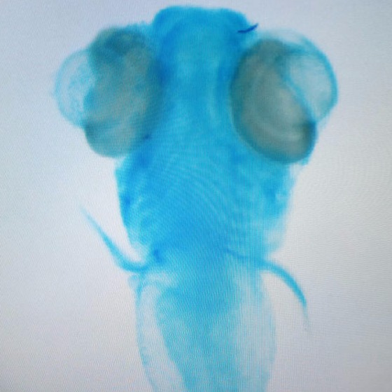
Zebrafish embryo, "where is my mind?" (author: Marco Spreafico)
Zebrafish embryo, on a Saturday night (author: Martina Betti)
Busy roads. Blood cells circulating into the blood vessels of a zebrafish larva (author: Matteo Ranucci/Alex Fantin)